CT scans reveal secrets of a golden mummy
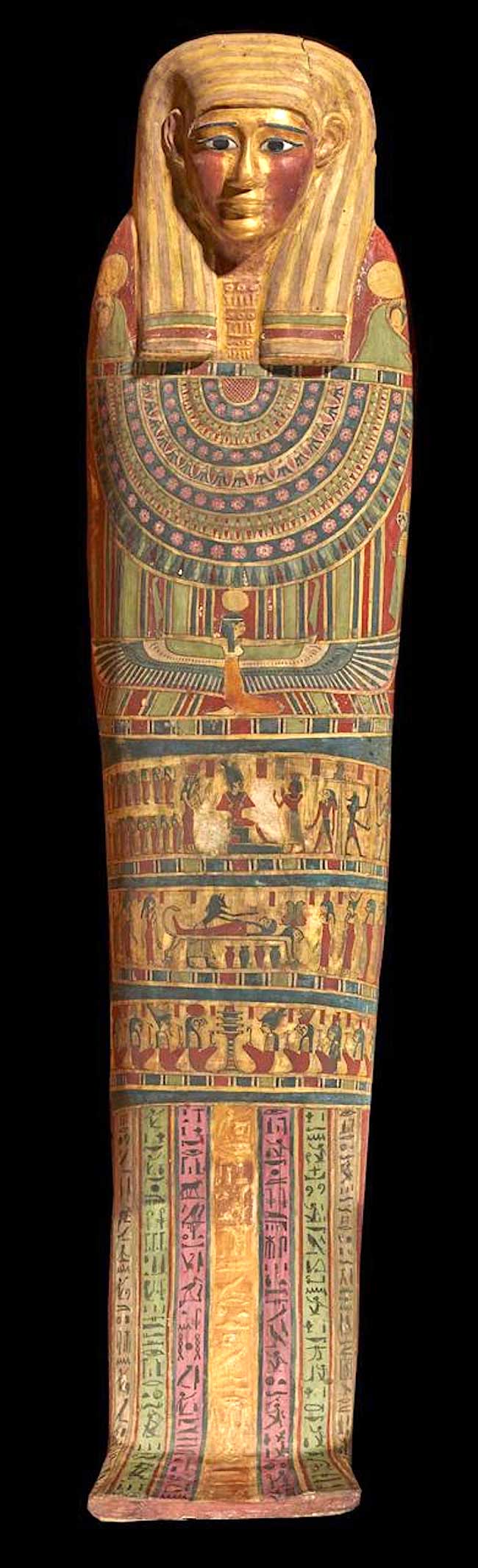
Computed tomography (CT) scans and 3D printing has revealed the secrets of the 2,300-year-old “Mummy of the Golden Boy,” enabling it to be displayed at the Egyptian Museum in Tahrir for the first time in more than a century.
The findings were revealed in a study published Tuesday in the Frontiers in Medicine journal by Dr. Sahar Saleem, professor of radiology at Cairo University.
The mummy was discovered completely wrapped in a tomb from the Ptolemaic period (circa 300 BC) in Edfu, in Aswan, in 1916 and moved to the museum’s basement in Cairo.
The mummy had been stored unexamined for over a century until it was recently studied for the first time by Saleem, in collaboration with Sabah Abdel-Razek, director general of the Egyptian Museum in Tahrir, and Mahmound El-Halwagy, former director of the museum.
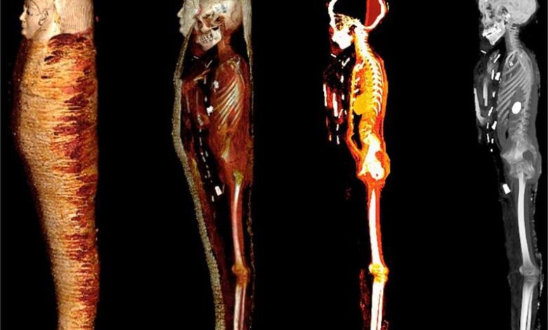
They used a computed tomography machine, along with advanced radiology, modern computer programs, and 3D printing to reveal the secrets of the mummy.
Their studies determined the mummy had been a 15-year-old boy, who had been elaborately mummified. His brain had been removed through the nostril, while his viscera were removed through a small incision in the lower abdomen. Packs and resins were inserted in the skull and body cavities. The embalmers were eager to keep the heart, which can be seen in the CT images inside the chest cavity, believing it to be spiritually important.
The CT scans also revealed that the mummy’s body was extensively decorated with 49 beautifully stylized amulets in three columns between the folds of the wrappings and inside the mummy’s body cavity. The amulets appear in 21 different shapes including the eye of Horus, the scarab and the amulet of the horizon (akhet), the placenta, the knot of Isis, the tow-feathers, and others.
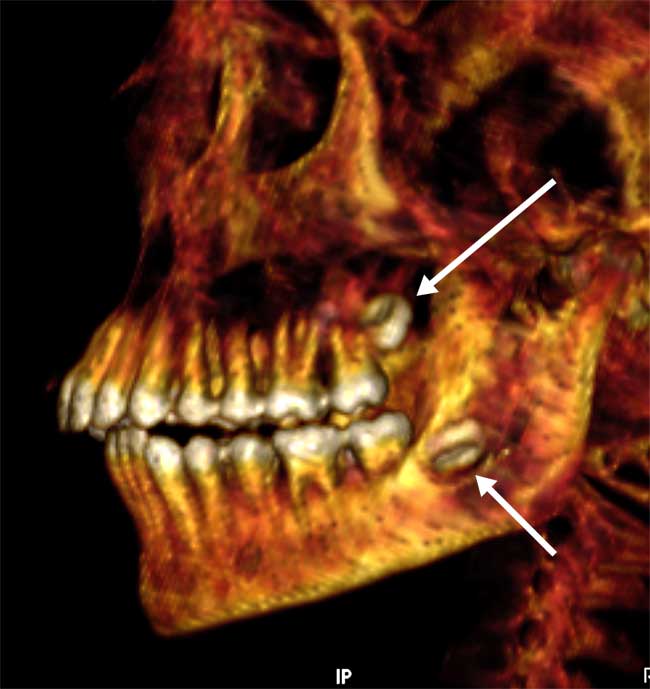
CT measurements revealed that 30 of these amulets are made of gold, while the others are made of stone or faience (a brightly coloured ceramic). As a result, the study provided a unique opportunity to learn about the different shapes and arrangements of amulets during mummification in a non-invasive manner.
The embalmers placed the amulets on the body to protect it and give it vitality in the afterlife. A gold amulet in the shape of a tongue was placed inside the mummy’s mouth so that he could speak in the afterlife. A two-finger amulet was placed below the trunk to protect the embalming incision. A large gold amulet of the heart scarab inside the mummy’s chest cavity was replicated using 3D printing.
Similarly, the CT scan enabled the boy’s face to be virtually unwrapped for the first time in 2,000 years.
According to Abdel-Razek, their studies shed light on social life in ancient Egypt thousands of years ago. It has provided a more in-depth understanding of ancient Egyptian beliefs and funerary rituals, as well as their technical skills in mummification and craftsmanship in the creation of amulets, masks, and decorations.
According to the study, the ancient Egyptians valued children so much that they provided them with remarkable funerary rituals to enable them to resurrect and join them in the afterlife. The mummy received lavish funeral rituals, including high-standard mummification with a gilded mask and several golden amulets, emphasizing the 15-year-old boy’s noble social status, with healthy teeth and bones and no signs of malnutrition.
The insights gained from the study supported the museum’s decision to move the mummy from its vault to a display in the main hall. The display of CT images next to the mummy contributes to a unique display that provides museum visitors with a special experience that facilitates their communication with the ancient Egyptian civilization.